Week of 26 January 2021

New IMMR feature: Second in a series of 3
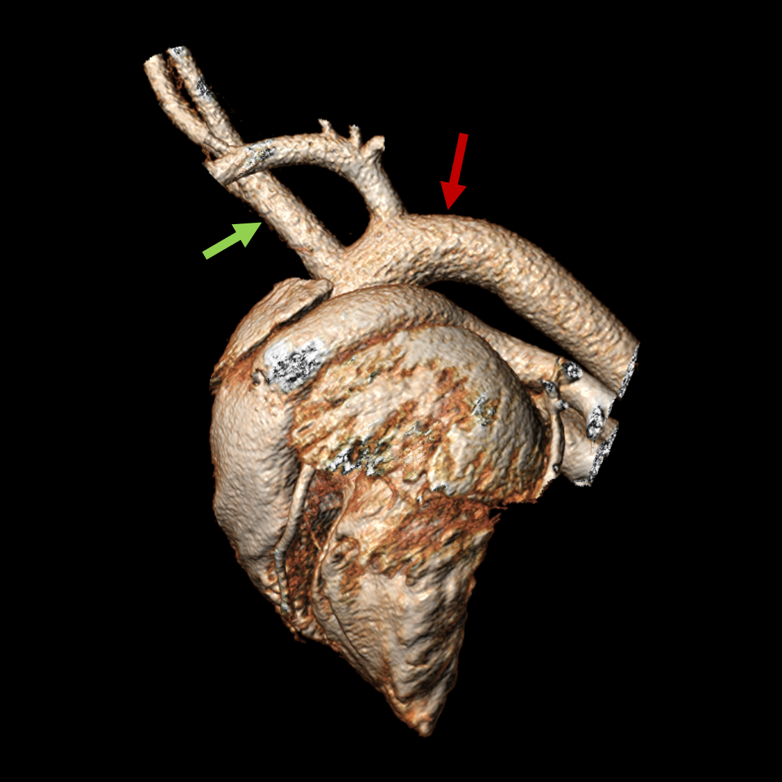
This is an image of a porcine (pig) aortic arch!
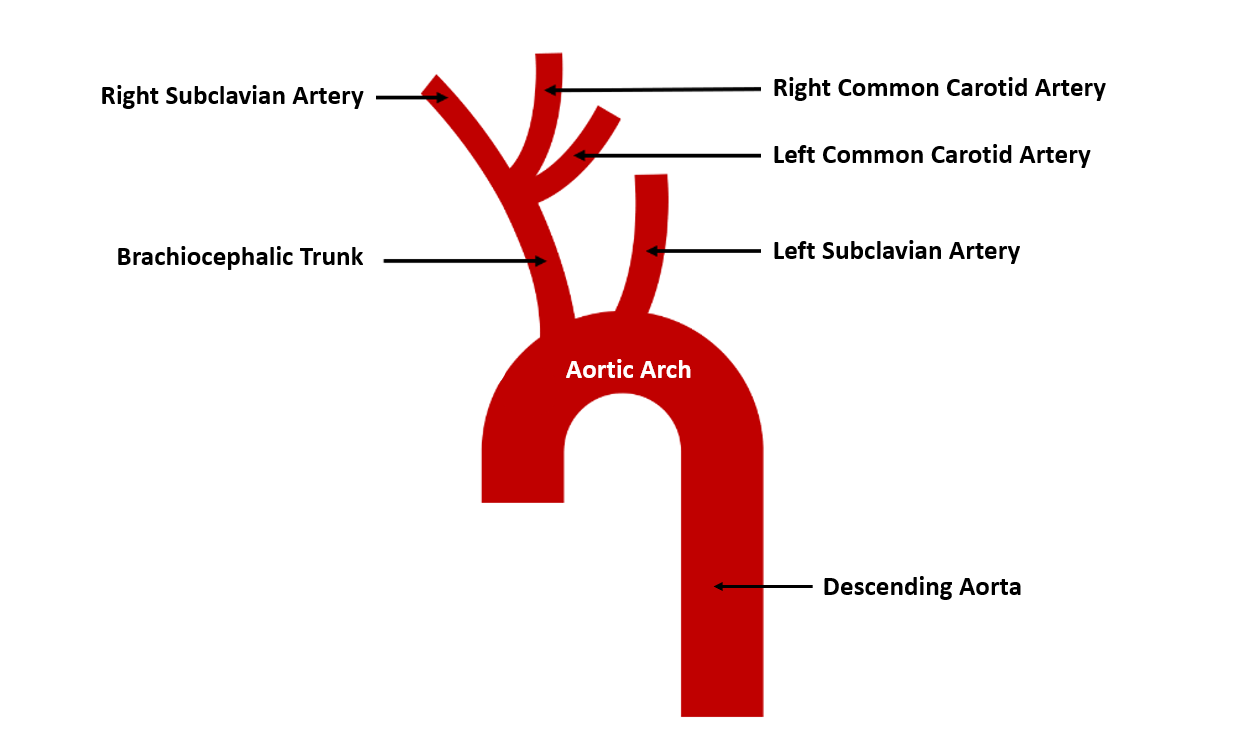
Here’s how we know: From the aortic arch (red arrow) emerge two branches, (i) the brachiocephalic trunk (green arrow) and (ii) the left subclavian artery. The brachiocephalic trunk gives rise to the right subclavian artery and the left and right common carotid arteries.
Pigs have emerged as a widely used model for surgical and interventional medical device evaluation, especially with respect to cardiac valve replacement and repair. Anatomic and physiologic similarities between pigs and humans make porcine species highly relevant animal models for translational research in cardiovascular medical device innovation. Understanding the differences between porcine and human anatomy is critical to guiding medical technology development and extrapolating observations in pig models to the expected safety and performance of medical devices in human patients.

This image is a 3D reconstruction of cardiovascular anatomy acquired using IMMR’s state-of-the-art 64-slice gated CT scanning system and 3mensio 3D reconstruction software.
Contact us to learn more and discuss your preclinical research and pathology needs.
Follow us on LinkedIn and don’t miss new images from our library that we post every Tuesday, when you’ll have another chance to recognize, identify or diagnose what is shown. You can also stay updated on some of the latest developments in Preclinical Science. Stay tuned!


