Week of 04 May 2021


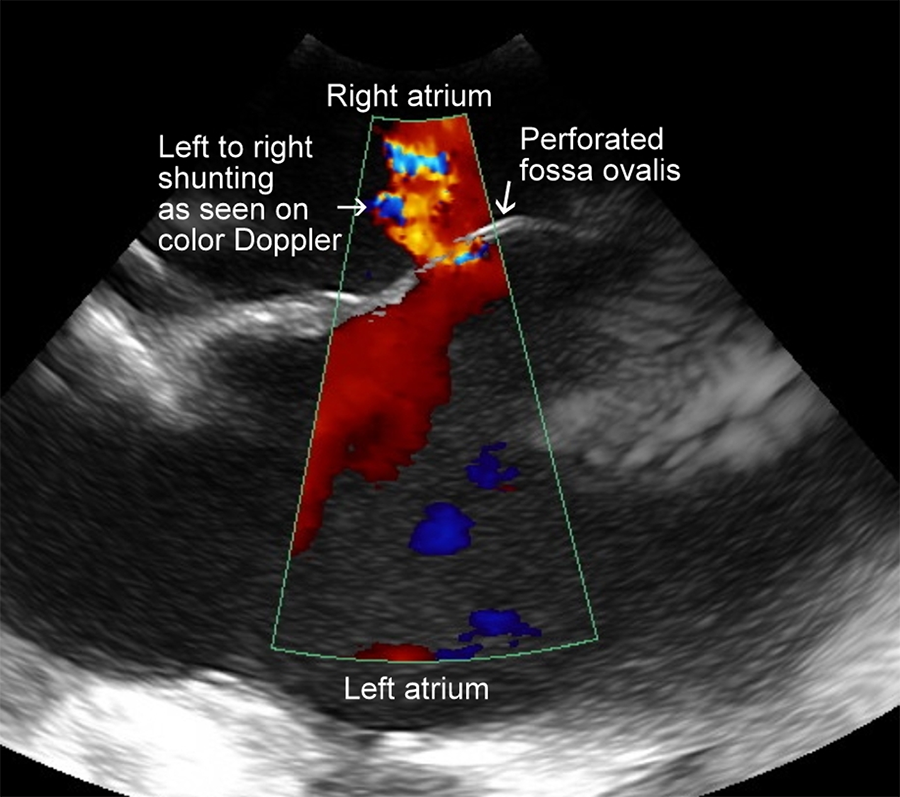
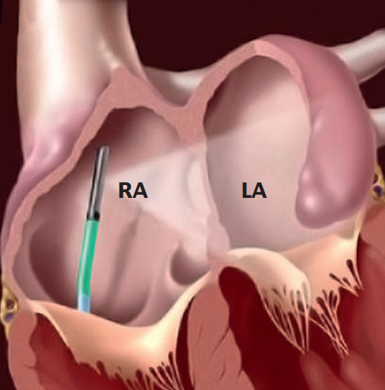
This image was obtained using IntraCardiac Echocardiography (ICE). For this imaging, a venous approach was used with the ultrasound probe positioned in the right atrium, looking through the interatrial septum, positioned at 12 o’clock in the accompanying diagram.
Interventional cardiologists routinely place the ICE probe in the right atrium to guide transseptal puncture and various percutaneous interventions, including left heart catheter ablation, left atrial appendage (LAA) closure/exclusion, and percutaneous mitral valvuloplasty. ICE provides direct visualization of the interatrial septum (visible at 12 o’clock on our ICE image) and is an invaluable tool to guide a safe transseptal puncture.1, 2
This crisp ICE image was acquired using IMMR’s state-of-the-art St Jude ViewFlex™ ICE 9-French catheter and Zonare ViewMate™ Z Intracardiac Ultrasound console.
Contact us to learn more and discuss your preclinical research and pathology needs.
Follow us on LinkedIn and don’t miss new images from our library that we post every Tuesday, when you’ll have another chance to recognize, identify or diagnose what is shown. You can also stay updated on some of the latest developments in Preclinical Science. Stay tuned!
1 Asirvatham SJ, Bruce CJ, Friedman PA: Advances in imaging for cardiac electrophysiology. Coron Artery Dis 2003;14:3–13.
2 Enriquez A, Saenz L, Rosso R, Silvestry F, Callans D, Marchlinski F, and Garcia F: Use of intracardiac echocardiography in interventional cardiology. Circulation 2018;137:2278–2294.
