Week of 14 September 2021

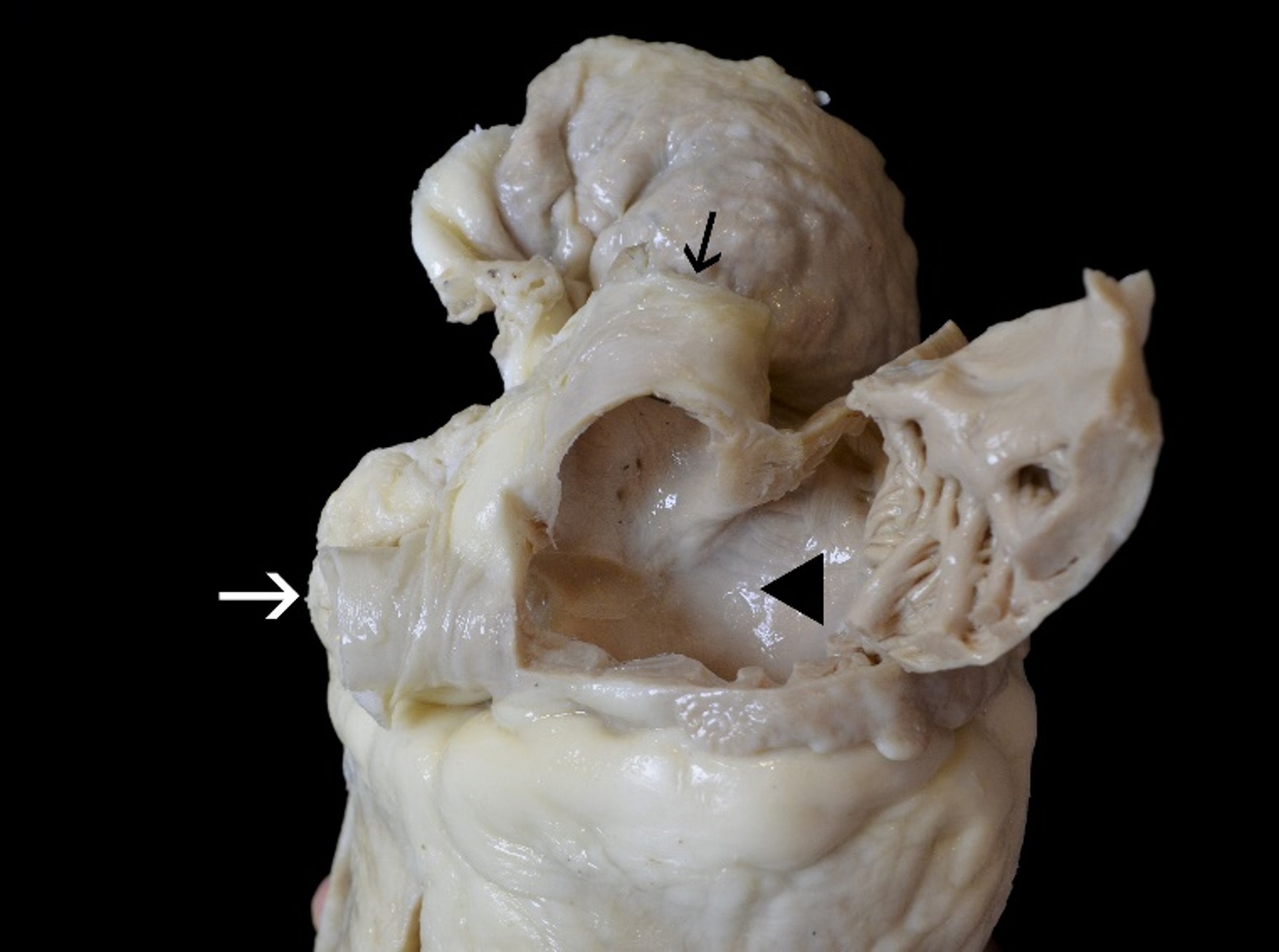
This is a view of the right atrium showing the two venae cavae: The superior vena cava (black arrow) and the inferior vena cava (white arrow)!
The right atrium receives deoxygenated blood from the superior vena cava (SVC) and the inferior vena cava (IVC). The right atrium leads into the right ventricle through the tricuspid valve.
The SVC is a large valveless venous channel formed by the union of the brachiocephalic veins. It receives blood from the upper half of the body (except the heart itself) and returns it to the right atrium.
The inferior vena cava (IVC) drains venous blood from the lower trunk, abdomen, pelvis and lower limbs to the right atrium.
Black arrow: Superior vena cava
White arrow: Inferior vena cava
Black triangle: Right Atrium
Plastination is a technique used to preserve organs and tissues in which the water and fat are replaced by plastic materials, yielding specimens that do not decay and retain most properties of the original sample.
This plastinated heart is one example from the comprehensive suite of Surgical Training and Pathology Services offered by IMMR’s in-house team of veterinary cardiac surgical specialists and Board-certified Veterinary Pathologists, in collaboration with the veterinary school of Maisons-Alfort, Paris.
Contact us to learn more and discuss your preclinical research and pathology needs.
Follow us on LinkedIn and don’t miss new images from our library that we post every Tuesday, when you’ll have another chance to recognize, identify or diagnose what is shown. You can also stay updated on some of the latest developments in Preclinical Science. Stay tuned!


