Week of 8 December 2020

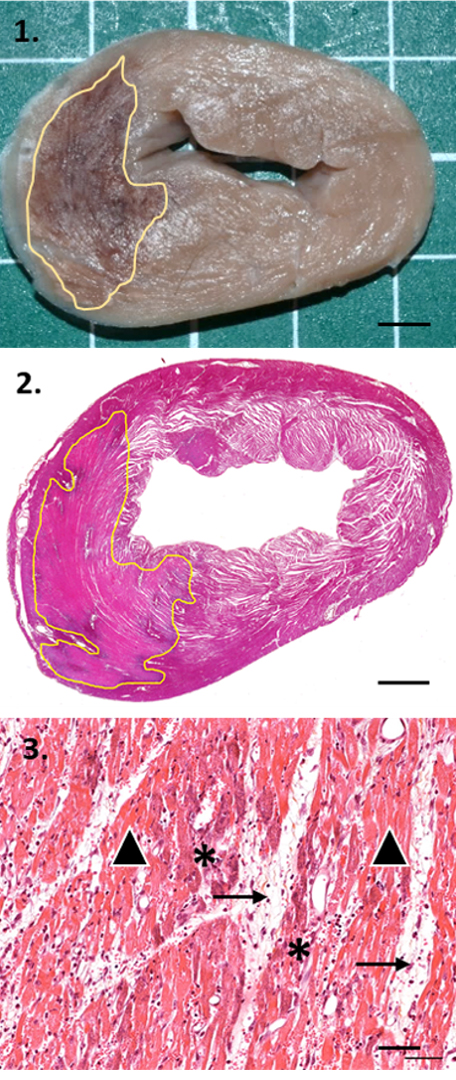
Myocardial infarcts (areas outlined in yellow) are observable in this ovine cardiac apex!
Using specialized histologic megacassettes to obtain very large samples (megasections), it is possible to analyze the whole cardiac apex and to offer an outstanding macro-micro correlation as well as a precise morphometric analysis of myocardial lesions.
1. Macroscopic view at trimming after fixation, scale bar: 0.5 cm.
2. Megasection of the cardiac apex. HE&S, original magnification: x1, scale bar: 0.5 cm.
3. Myocardial infarct. HE&S, original magnification: x20, scale bar: 50 µm.
Yellow line: Delimitation of myocardial infarcts
Black arrow: Degenerating/necrotic myocardial fibers
Black arrowhead: Hemorrhage and edema
*Mineral deposits
This histopathology image is one example from the comprehensive suite of Pathology Services offered by IMMR’s in-house team of Board-certified Veterinary Pathologists.
Contact us to learn more and discuss your preclinical research and pathology evaluation needs.
Follow us on LinkedIn and don’t miss new images from our library that we post every Tuesday, when you’ll have another chance to recognize, identify or diagnose what is shown. You can also stay updated on some of the latest developments in Preclinical Science. Stay tuned!


