Week of 17 November 2020
answer

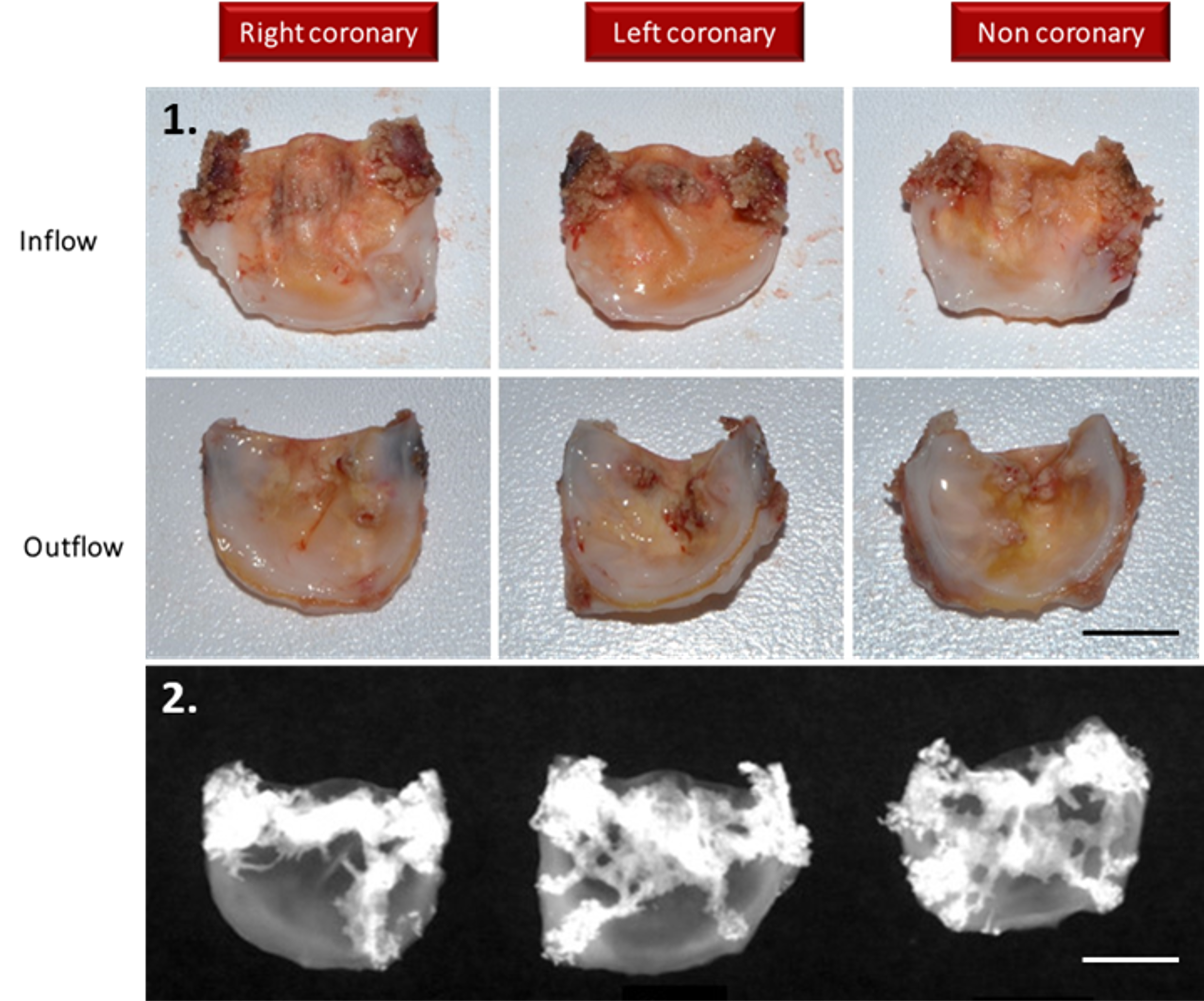
These bioprosthetic leaflets are distorted by mineral deposits. They are from a valve that was explanted from an ovine at 150 days after implantation.
Mineral deposits appear as firm, brittle, white-yellowish deposits within the leaflet or at the leaflet surface at macroscopic examination. They are highly radio-opaque on high resolution X-ray examination (Faxitron®) of the excised cusps. Mineral depositions are one of the most common complications of bioprosthetic cardiac valve implantation and may lead to the necessity of replacement with a new valve.
- Macroscopic view at trimming after fixation, scale bar: 0.5 cm.
- Faxitron® imaging, scale bar: 0.5 cm.
Faxitron imaging is one example from the comprehensive suite of Pathology Services offered by IMMR’s in-house team of Board-certified Veterinary Pathologists.
Contact us to learn more and discuss your preclinical research and pathology needs.


