Week of 05 October 2021

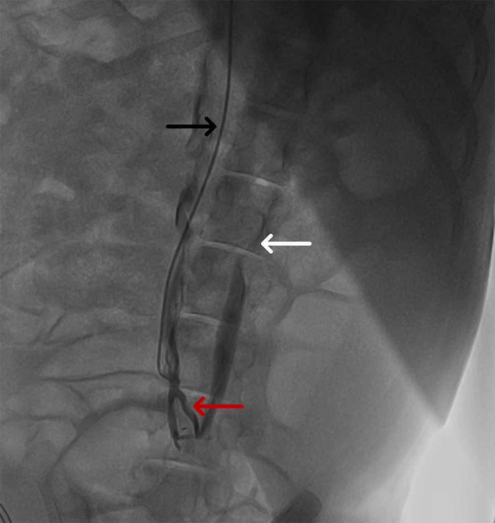
Two conjoined caudal vena cavae!
In preparation for evaluation of an investigational thrombectomy device, a catheter was inserted via the right internal jugular vein and advanced inferiorly. The injection of a contrast agent revealed duplicated, conjoined caudal vena cavae.
Black arrow: Right vena cava.
White arrow: Left vena cava.
Red arrow: Communication
In mammals, duplication of the caudal vena cava has been described previously and in humans this may be linked with elevated risk of thromboembolic events.1
Large animal models, including pathologic models of central deep venous thrombosis of the vena cava and/or iliac veins, are valuable in the development and evaluation of new vascular interventional products including stents, filters, thrombectomy catheters and other life-saving medical devices.
Contact us to learn more and discuss your preclinical research and pathology needs.
Follow us on LinkedIn and don’t miss new images from our library that we post every Month, when you’ll have another chance to recognize, identify or diagnose what is shown. You can also stay updated on some of the latest developments in Preclinical Science. Stay tuned!
1. Eldefrawy A et al: Anomalies of the inferior vena cava and renal veins and implications for renal surgery. Cent European J Urol 2011; 64(1):4-8.


